Anatomy Of Chest And Stomach / Anatomia Y Fisiologia Carnet D Anatomie Thorax Abdomen Pelvis Organ Human Anatomy Abdomen Anatomy Png Pngwing : It is part of the digestive system, which extends from the mouth to the anus.
Anatomy Of Chest And Stomach / Anatomia Y Fisiologia Carnet D Anatomie Thorax Abdomen Pelvis Organ Human Anatomy Abdomen Anatomy Png Pngwing : It is part of the digestive system, which extends from the mouth to the anus.. The main function of the stomach involves mechanical and chemical digestion of ingested food. The wall of the stomach is made of the same four layers as most of the rest of the alimentary canal, but with adaptations to the mucosa and muscularis for the unique functions of this organ. There are two sphincters of the stomach, located at each orifice. Stomach, saclike expansion of the digestive system, between the esophagus and the small intestine; You may also find triceps, lateral head brachialis, biceps brachii, latissimus dorsi, deltoid, acromion anatomynote.com found chest muscle anatomy from plenty of anatomical pictures on the internet.
It is part of the digestive system, which extends from the mouth to the anus. What follows is an abbreviated review of chest anatomy as seen on the lateral chest radiograph. The normal microscopic and gross morphologic features of the stomach are described. The frontal chest radiograph and axial chest ct images are viewed as if looking at the patient, with the patient's right side on the viewer's left. Nervous tissue in the submucosa monitors the contents of the stomach and controls smooth muscle contraction and secretion of digestive substances.
Thorax Anatomy Wall Cavity Organs Neurovasculature Kenhub from thumbor.kenhub.com Note that the stomach bubble is under the left hemidiaphragm. Swensen fund for innovation in teaching. The stomach is a muscular organ located on the left side of the upper abdomen. The embryologic and anatomic basis of modern surgery. The wall of the stomach is structurally similar to other parts of the digestive tube, with the exception that the stomach has an extra oblique layer of smooth muscle inside the circular layer, which aids in the. This type of ct scan uses a lower radiation level than a conventional. The thoracic cage is the bony. To duodenum regions cardiac fundus body pyloric.
Between oesophagus (ostium cardiacum) and small intestine (ostium pyloricum) syntopy (syntopia):
It lies in the epigastric, umbilical, and left hypochondriac regions of. This page provides an overview of the chest muscle group. The chest wall is supplied by the posterior intercostal arteries arising from the aorta, the internal thoracic and the highest intercostals given off the subclavian artery, and the branches of the axillary artery (fig. The stomach is a muscular organ located on the left side of the upper abdomen. Decker ga, plessis d du. Anterior abdominal wall, diaphragm and liver. The trachea (windpipe) conducts inhaled air into the lungs the digestive tract comprises several organs that are connected and interdependent. Learn about the anatomy and physiology of the stomach. The upper portion of the trunk between the neck and the abdomen. Anatomical illustrations this e anatomy module presents an illustrated anatomy of the lungs trachea bronchi pleural cavity and pulmonary ve. Lee mcgregor's synopsis of surgcial anatomy. The hollow abdominal organs include the stomach, small. You may also find triceps, lateral head brachialis, biceps brachii, latissimus dorsi, deltoid, acromion anatomynote.com found chest muscle anatomy from plenty of anatomical pictures on the internet.
Between oesophagus (ostium cardiacum) and small intestine (ostium pyloricum) syntopy (syntopia): The wall of the stomach is made of the same four layers as most of the rest of the alimentary canal, but with adaptations to the mucosa and muscularis for the unique functions of this organ. The stomach lies within the superior aspect of the abdomen. Anatomy of the stomach, gallbladder, and pancreas. You may also find triceps, lateral head brachialis, biceps brachii, latissimus dorsi, deltoid, acromion anatomynote.com found chest muscle anatomy from plenty of anatomical pictures on the internet.
Clippedonissuu From Anatomy Drawing Conor Power Shoulder Anatomy Shoulder Muscle Anatomy Chest Muscles from i.pinimg.com The stomach receives food from the esophagus. Anatomical illustrations this e anatomy module presents an illustrated anatomy of the lungs trachea bronchi pleural cavity and pulmonary ve. Anatomy of the chest and the lungs: It lies in the epigastric, umbilical, and left hypochondriac regions of. The user can browse between different groups of images using the series tab The chest anatomy includes the pectoralis major, pectoralis minor and the serratus anterior. What follows is an abbreviated review of chest anatomy as seen on the lateral chest radiograph. The stomach is the most dilated part of the digestive tube, and is situated between the end of the esophagus and the beginning of the small intestine.
The main function of the stomach involves mechanical and chemical digestion of ingested food.
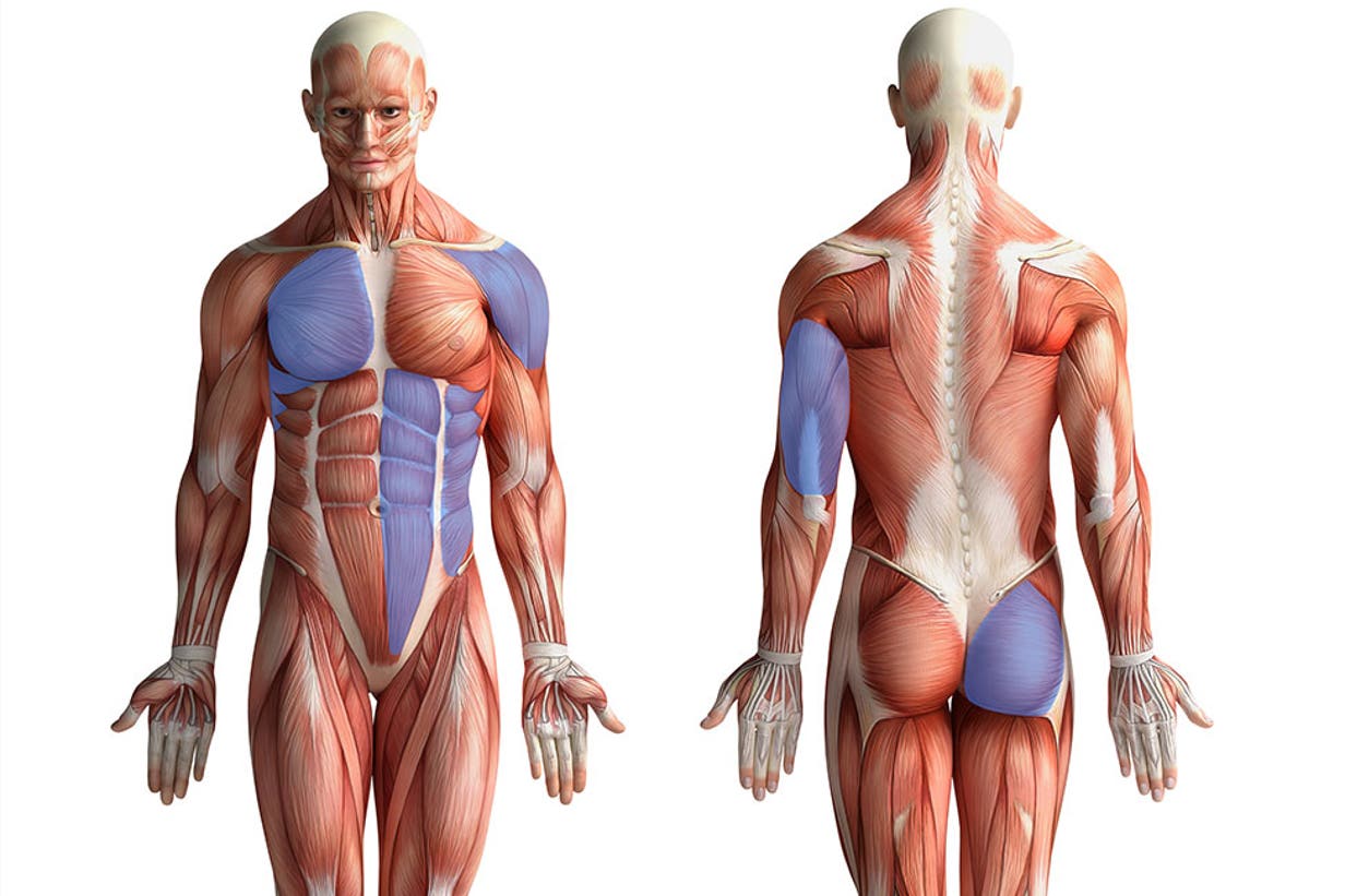
The hollow abdominal organs include the stomach, small. There are two sphincters of the stomach, located at each orifice. Find out more about the individual muscles within the chest anatomy by clicking their respective links throughout this page. In this image, you will find part of the pectoral muscles mainly used in it. Nervous tissue in the submucosa monitors the contents of the stomach and controls smooth muscle contraction and secretion of digestive substances. The embryologic and anatomic basis of modern surgery. What happens to fats, carbs & protein in foods. It is located in the anterior portion of the abdominal cavity in most the stomach serves as a temporary receptacle for the storage and mechanical distribution of food before it is passed into the intestine. False flatter chest fluctuating fluoride. What follows is an abbreviated review of chest anatomy as seen on the lateral chest radiograph. The stomach is a muscular organ located on the left side of the upper abdomen. The trachea (windpipe) conducts inhaled air into the lungs the digestive tract comprises several organs that are connected and interdependent. Living anatomy of the chest for 1st year medical students original version compiled by dr.
The trachea (windpipe) conducts inhaled air into the lungs the digestive tract comprises several organs that are connected and interdependent. This video is about the division of the abdominal cavity and the anatomy of the stomach. What are parts of the stomach & the stomach anatomy. The hollow abdominal organs include the stomach, small. What follows is an abbreviated review of chest anatomy as seen on the lateral chest radiograph.
Pushups Way More Than Chest Training from images.contentstack.io Decker ga, plessis d du. Find out more about the individual muscles within the chest anatomy by clicking their respective links throughout this page. Lee mcgregor's synopsis of surgcial anatomy. The stomach lies within the superior aspect of the abdomen. Emphasis is given to mucosal anatomy and the recognition of minor alterations seen in disease that may be identified on an endoscopic biopsy. False flatter chest fluctuating fluoride. The stomach is the most dilated part of the digestive tube, and is situated between the end of the esophagus and the beginning of the small intestine. They control the passage of material entering and a hiatus hernia occurs when a part of the stomach protrudes into the chest through the oesophageal.
Protective framework for parts of the chest involved with brea… chest or thorax.
The stomach lies within the superior aspect of the abdomen. It lies in the epigastric, umbilical, and left hypochondriac regions of. This type of ct scan uses a lower radiation level than a conventional. In lungs,stomach fat soft tissue which include muscle. There are two sphincters of the stomach, located at each orifice. The embryologic and anatomic basis of modern surgery. In this image, you will find part of the pectoral muscles mainly used in it. The stomach consists of several important anatomical parts. This video is about the division of the abdominal cavity and the anatomy of the stomach. Learn about the anatomy and physiology of the stomach. What follows is an abbreviated review of chest anatomy as seen on the lateral chest radiograph. The chest anatomy includes the pectoralis major, pectoralis minor and the serratus anterior. Note that the stomach bubble is under the left hemidiaphragm.
The stomach consists of several important anatomical parts anatomy of chest. To duodenum regions cardiac fundus body pyloric.


0 Komentar